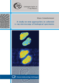
The use of coherent x rays for microscopic imaging has seen a rapid and ongoing development within the past decade, driven by an increasing availability of highly brilliant and coherent sources worldwide. Accordingly, novel methods have been developed, which replace the microscope‘s objective lens by a numerical reconstruction scheme. The aim of the present work is to study how very recent experimental and algorithmic developments in the field can be implemented towards a highly sensitive and fully quantitative microscopy method for imaging of biological cells. To this end, different experimental approaches are studied, based on coherent far-field as well as near-field diffraction. At first, an application of the novel ptychographic imaging method to single biological cells is presented. In particular, it is demonstrated how weakly scattering biological specimens can be imaged with fully quantitative density contrast. Alongside, a sueccessful extension of the method towards soft x-ray energies is described.In the second part of the work it is shown how x-ray waveguides can be used as a point source for propagation-based microscopy of single cells in the hard x-ray regime. The specifically devised iterative reconstruction scheme allows for full quantitativity and high sensitivity and thus enables an application to single biological cells. The work contains a thorough introduction into the x-ray optical methods applied and aims at a useful and self-contained overview on aspects of signal and Fourier theory relevant for the used numerical propagation schemes.
Publikationstyp: Hochschulschrift
Sparte: Universitätsverlag
Sprache: Englisch


