

Investigating Cellular Nanoscale with X-rays
From Proteins to Networks
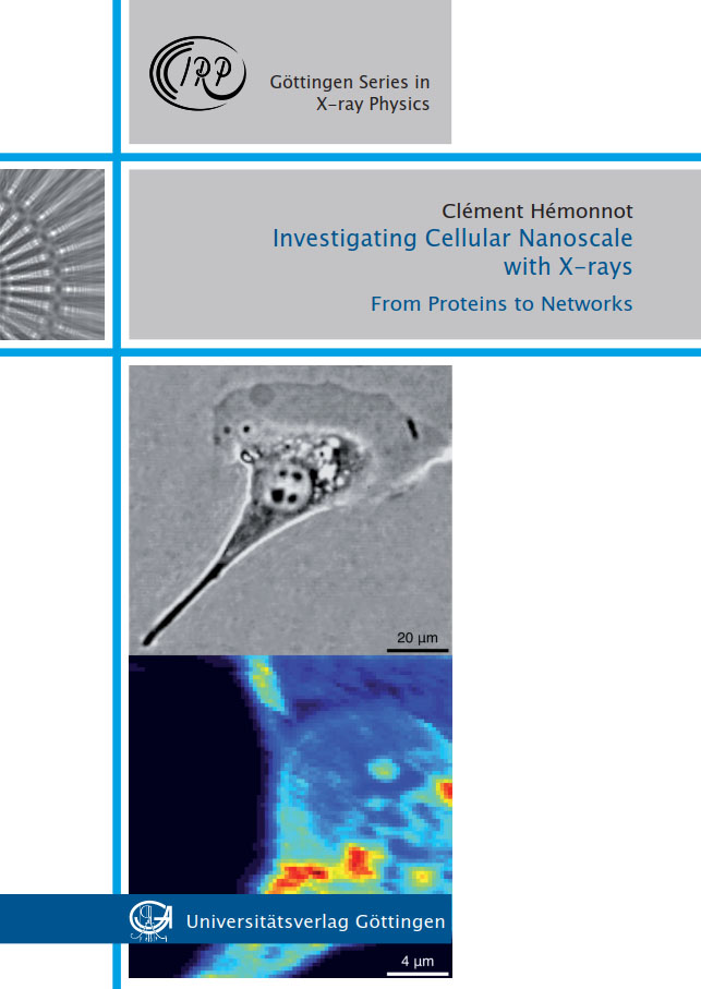
The advances and technical improvements of X-ray imaging techniques, taking advantage of X-ray focussing optics and high intensity synchrotron sources, nowadays allow for the use of X-rays to probe the cellular nanoscale. Importantly, X-rays permit thick samples to be imaged without sectioning or slicing. In this work, two macromolecules, namely keratin intermediate filament (IF) proteins and DNA, both essential components of cells, were studied by X-ray techniques. Keratin IF proteins make up an integral part of the cytoskeleton of epithelial cells and form a dense intracellular network of bundles. This network is built from monomers in a hierarchical fashion. Thus, the keratin structure formation spans a large range of length scales from a few nanometres (monomers) to micrometres (networks). Here, keratin was studied at three different scales: i) filaments, ii) bundles and iii) networks. Solution small-angle X-ray scattering revealed distinct structural and organisational characteristics of these highly charged polyelectrolyte filaments, such as increasing radius with increasing salt concentration and spatial accumulation of ions depending on the salt concentration. The results are quantified by employing advanced modelling of keratin IFs by a core cylinder fl anked with Gaussian chains. Scanning micro- diffraction was used to study keratin at the bundle scale. Very different morphologies of keratin bundles were observed at different salt conditions. At the network scale, new imaging approaches and analyses were applied to the study of whole cells. Ptychography and scanning X-ray nano-diffraction imaging were performed on the same cells, allowing for high resolution in real and reciprocal space, thereby revealing the internal structure of these networks. By using a fitting routine based on simulations of IFs packed on a hexagonal lattice, the radius of each fi lament and distance between fi laments were retrieved. In mammalian cells, each nucleus contains 2 nm-thick DNA double helices with a total length of about 2 m. The DNA strands are packed in a highly hierarchical manner into individual chromosomes. DNA was studied in intact cells by visible light microscopy and scanning X-ray nano-diffraction, unveiling the compaction und decompaction of DNA during the cell cycle. Thus, we obtained information on the aggregation state of the nuclear DNA at a real space resolution on the order of few hundreds nm. To exploit to the reciprocal space information, individual diffraction patterns were analysed according to a generalised Porod’s law at a resolution down to 10 nm. We were able to distinguish nucleoli, heterochromatin and euchromatin in the nuclei and follow the compaction and decompaction during the cell division cycle.
Publikationstyp: Hochschulschrift
Sparte: Universitätsverlag
Sprache: Englisch